Art in Science
Our laboratory values the role of art in science. Art is not only an important tool used to aid in the visualization of concepts - something crucial when working with creation of new platforms - but it is also critical to enriching creativity and ownership in the work our students do.
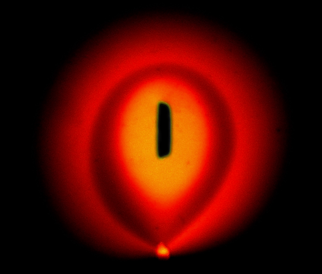
Eye of Sauron

By: Nadia Ostromohov
A fluorescence image showing concentration gradients inside an electrokinetic flow confinement of fluorescein, created using the electrokinetic scanning probe (ESP), through a combination of electroosmotic flow (EOF) and electrophoresis.
Experimental visualization of electroosmotic flow (EOF) dipole streamlines generated by different gate electrode geometries in a microfluidic chamber. The gate electrodes consist of a conductive layer insulated from the electrolyte in the chamber by a thin dielectric. The modulation of the potential on the gate electrodes in-sync with the electric field inside the chamber results in net EOF on top of the electrode. The superposition of streamlines generated by different gate electrodes could be used as flow patterning method, which allows fluid manipulation without use of any physical walls.
Dipole Music: Stop, Play, Record

By: Federico Paratore & Vesna Bacheva
Symmetric Microfluidic Probe Design


By: Aditya Kashyap and Nadia Enrriquez
The image on the left shows hierarchical Hydrodynamic Flow Confinements (hHFCs) designed for long-term cell exposure and maintenance. The image on the right shows the design, which includes a bubble trap and inertial microfluidic design principles to allow for longer experiments.
"Immunohistochemistry (IHC) assay in its current form is not very quantitative, because there is non-linear light capture by the colored precipitate. This is akin to holding an umbrella in the rain while standing in a puddle and trying to count the number of water droplets in the puddle, all the while wearing sun glasses."
IHC: Counting Rain Drops in a Puddle

By: Anna Fomitcheva Khartchenko and Aditya Kashyap
Bubble vs Flow a Microfluidic Odessey

By: Lorenzo Petrini
The images depict a flow of fluorescent beads inside a microfluidic chip, dramatically distorted by the presence of air bubbles.
Transition between flow patterns in a microfluidic flow cell with an array of apertures for the dynamic reconfiguration of flow trajectories. By injection an aspiration of buffer liquid from an 4×4 array of apertures, the flow of miscible processing liquids can be confined to so-called “virtual channels”. These virtual channels can be formed and reconfigured within less than 20s.
The photograph shows the flow of two different liquids (transparent and red) in several virtual channels (height 0.1mm, width about 2mm), confined by the flow of a blue buffer liquid.
Virtual Channels

By: David Taylor
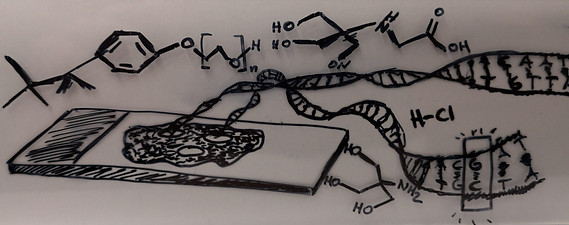
Dragging DNA out of Tissues

By: Lorenzo Petrini
A method for spatially resolved genetic analysis of FFPE cell block and tissue sections is presented. This involves local sampling with hydrodynamic flow confinement of a lysis buffer, electrokinetic purification of nucleic acids from the lysate, and detection of single point mutations through amplification and sequencing.